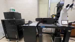
• Provides a state-of-the-art facility for catering to the needs of researchers with diverse imaging capabilities
• Conducts national level and local training programmes to develop skilled manpower in Bio imaging
NCCS Bio-Imaging Facility Instruments:
Pune Biocluster Project funded:
1. Zeiss LSM880 Confocal Microscope with AiryScan and ELYRA P.1
• This imaging system comprises of AxioObserver 7 microscope with AiryScan and Elyra P.1.
• Airyscan is a 32-channel GaAsP-PMT area detector that collects a pinhole-plane image at every scan position. Each detector element functions as a single, very small pinhole.
• ELYRA P.1 localizes small structures and even single molecules achieving resolutions of 20nm laterally and 50nm axially
• Objectives: 10x, 20x, 40x (oil), 63x (oil), 100x (oil) Alpha Planapochromat
• Lasers: For confocal- 405nm, 488nm, 458nm, 514nm, 561nm, 594nm, 633nm; For Elyra P.1- 405nm, 488nm, 561nm, 640nm
2. Olympus SPIN SR 10 - Spinning Disk High Resolution Microscope
• This Spinning disc confocal microscope is equipped with YOKOGAWA W1- SoRa scan-unit.
• It can attain high resolution minimum 250 nm (XY) and 800 nm in Z. Super-resolution mode gives XY resolution of 120nm and Z of 540nm approximately.
• The system has 2 high-speed cameras for simultaneous imaging of atleast two dyes.
• Objectives: 10X to 100X.
• Lasers: 405nm, 488nm, 514nm, 561nm, 640nm
3. Olympus Multiphoton microscope
• Olympus FVMPE-RS multiphoton laser scanning microscope is available for deep tissue imaging of fixed samples and live whole animals.
• This system features a tunable infrared laser (MaiTai, 680~1040nm) to excite multiple fluorescence dyes optimally for precise multispectral imaging.
• It also provides resonance scanning mode for high speed imaging and laser light stimulation for photomanipulation of fluorophores such as FRAP and optogenetics.
• With dedicated multiphoton-specific objectives (10x and 25x water dipping), these features allow users to image the large samples (up to 8 mm thickness) in multicolor 4D.
• Multicolor Imaging filter cubes: FVG (violet & green) and FGR (green & red) are available.
• The system is equipped with two GaAsP PMTs and also has 3D deconvolution and rendering functions.

4. Leica Stellaris 8 STED Microscope
• This microscope is a tool for advanced imaging applications, offering high-resolution, multicolor, and live-cell imaging capabilities.
• This imaging system combines confocal microscopy with Stimulated Emission Depletion (STED) super- resolution capabilities with sub-30 nm resolution, having Tunable excitation (440–790 nm).
• STED microscopy can achieve a lateral resolution of ~30–50 nm and axial resolution of ~120–150 nm.
• Some unique features are Power HyD detectors with high photon detection efficiency (HyD S & HyD X), real-time analysis of molecular dynamics by Fluorescence Correlation Spectroscopy (FCS) and TauSense.
• It also has FAst Lifetime CONtrast (FALCON), a FLIM solution for fast acquisition which combines STED with fluorescence lifetime imaging (FLIM) to separate fluorophores based on their lifetimes.
• LIGHTNING Provides enhanced resolution and contrast in real- time imaging, capturing fine structural details.
• Objectives: 10x-100x and a special 93x STED objective.
• Lasers: 405nm laser, Tunable White Light Laser (WLL) 440-790 nm, 589nm and 775nm Depletion Lasers.
5. Nikon Structured Illumination Microscope/ X-Light V3 and DeepSIM system:
• X-Light V3 system is a spinning disk confocal system offering precise 3D sectioning at the highest speed and largest field of view maintaining uniform illumination.
• The DeepSIM module allows researchers to seamlessly enhance their imaging capabilities from widefield to confocal to super-resolution.
• The 2D Lattice SIM technology behind DeepSIM enhances the illumination efficiency and homogeneity with minimal phototoxicity in live cell imaging experiments.
• Objectives: 10x, 20x, 40x, 40x Silicon, 60x oil and SR HP Apo 100x oil.
• Lasers: 405nm, 488nm, 555nm, 640nm
User Charges for Zeiss880 and SpinSR10:
Charges based on number of slots used where one hour is considered as one slot.
Users from other educational institutions: Rs.2000/- per slot + 18% GST.
Users from Industry: Rs.3000/- per slot + 18% GST.
Download Sample Request Form: Download form from here
Note:User charges for Leica Stellaris8 STED, Olympus Multiphoton and Nikon V3-DeepSIM are to be finalized.